Useful for
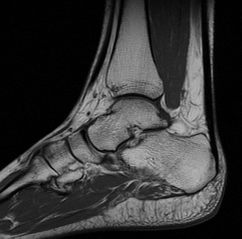
bone marrow assessment and evaluate general ankle anatomy.
Normal
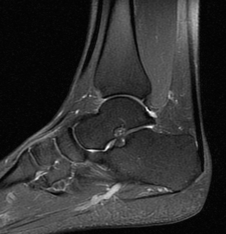
2- Sagittal "Fat Sat" PD spin
echo (SE)
Best sequence
to assess the Achilles tendon. The other ankle tendons, including the
plantar fascia, are also well visualized.
Normal
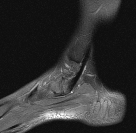
Posterior
tibialis tendon attachment site is clearly assessed using this sequence (Accessory
Navicular Bone Syndrome)
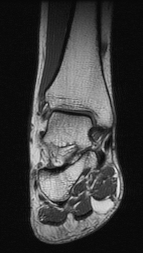
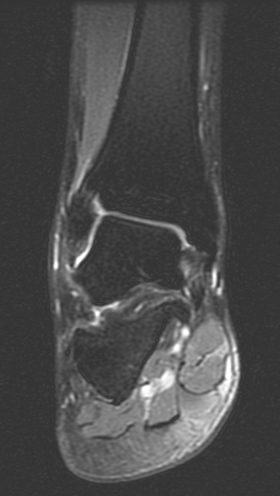
3- Coronal T1 spin echo (SE)
General
anatomic assessment of the ankle. The tibiotalar articulation, as well as
the tibiotalar ligaments are assessed. Assessment of the bone marrow on T1
is important also.
Normal
4- Coronal "Fat Sat" PD spin
echo (SE)
Ankle tendons
including the tibiotalar ligaments are well assessed using this
sequence.
Normal
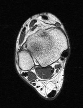
5- Axial T1 spin echo (SE)
Ankle
tendons, muscles, general ankle anatomy all re-evaluated with this
sequence. Assessment of the bone marrow on T1 is important as mentioned
above.
Normal
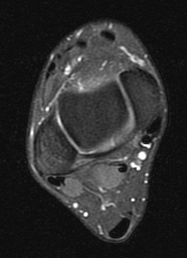
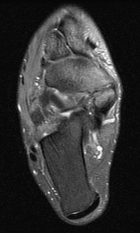
6- Axial "Fat Sat" PD spin
echo (SE)
Ankle tendons
are best evaluated with this sequence. Similarly, the talofibular and
tibiotalar ligaments also well visualized.
Normal
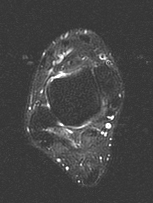
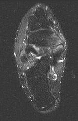
7- Axial STIR
This sequence
is the best to screen the bones to make sure there is not a subtle bone
edema or fluid collections that is not visible on other sequences.
Normal.
Edema involving the
navicular and accessory navicular bones better visualized on STIR compared
to fat sat PD sequence (Accessory Navicular Bone Syndrome).