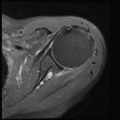
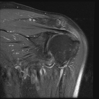
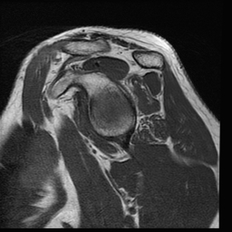
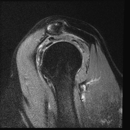
Particularly useful
for assessment of the long head of the biceps tendon and subscapularis
tendon. Glenohumeral ligaments, glenoid labrum and acramioclavicular joint
are also assessed.
Normal
Long head of the
biceps tendon demonstrates high signal and distortion, consistent with a
tendon tear.
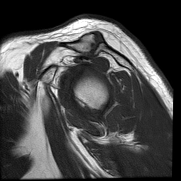
Bony bankart
lesion
Small
paralabral cyst
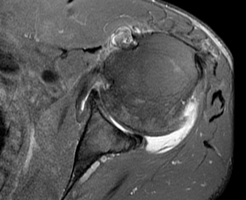
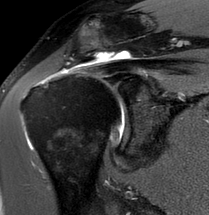
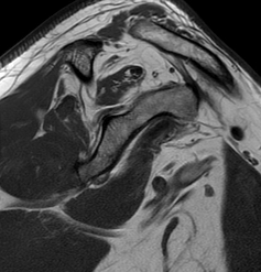
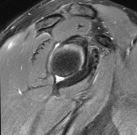
2- Coronal Fat-Sat PD
spin echo (SE):
Supraspinatus
tendons and acromioclavicular joint are assessed. Superior labrum is also
visualized well on this sequence.
Normal
There
is complete tear of the supraspinatus tendon, with some fluid in the
subacromian subdeltoid bursa as expected.
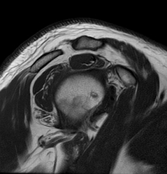
There
is tendinosis - tendinopathy involving the rotator cuff. There is also
irregularity and signal abnormality in the superior labrum, consistent
with a SLAP lesion.
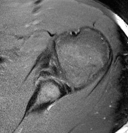
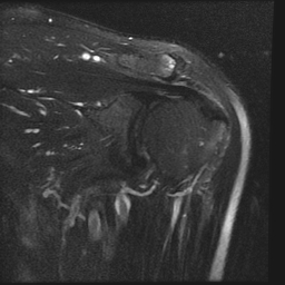
3- Coronal Fat-Sat T2
spin echo (SE)
Particularly
useful to determine if the signal abnormalities in the supraspinatus
tendons and superior labrum is truly due to degeneration or tear, and also
to see if there is any fluid in the subacromian and subdeltoid bursa
(which would be due to bursitis or rotator cuff tear). Bone edema like
abnormalities also easily appreciated.
Normal
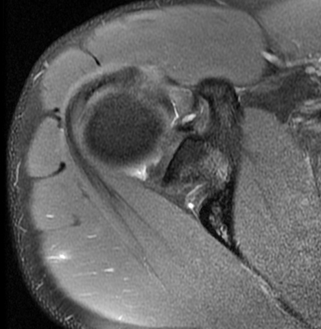
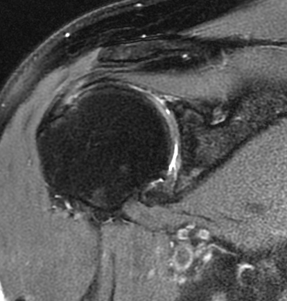
4- Sagittal T1 spin
echo (SE)
T1 sequence
is useful to assess the bone marrow. Bone marrow replacing conditions are
best evaluated with this sequence. Supraspinatus tendon impingement is
also easily appreciated.
Normal
Hypertrophic
degenerative acromioclaicular joint clearly compressing the supraspinatus
tendon.
Complete rotator
cuff tear with typical atrophic change in the supraspinatus tendon - best
appreciated on sagittal T1 weighted images.
Quadrilateral
space syndrome
5- Sagittal Fat-Sat PD
spin echo (SE)
Useful to
confirm findings appreciated on coronal plane and also useful for further
assessment of the biceps tendon and its superior labral attachment.